On-Demand AMD Histology Slides


.png)

IHC: Fit-For-Purpose
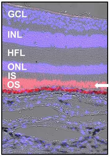
Our slides are designed to be used in immunohistochemistry experiments right out of the box. Photomicrograph shows fluorescence-labeled rhodopsin (white arrow), overlaid with differential interference contrast image. Localize your target of interest with confidence in these expertly prepared slides.
Human donor eyes were collected, characterized, and prepared for immunohistochemistry by J.D. Messinger DC (Curcio lab since 2007). All were preserved ≤ 6 hours from death by immersion in 4% paraformaldehyde, 0.1 M phosphate buffer after removal of cornea/ lens/ iris (stored in 1% paraformaldehyde). Ex vivo fundus imaging includes OCT, color, near-infrared reflectance, and autofluorescence. Cryosections (2/slide) are 12 µm thick, ≥ 20 mm long, parallel to horizontal meridian, through fovea and perifovea, collected on SuperFrost slides (positively charged, borosilicate glass, 1 mm thick, subbed with poly-L-lysine). Sample size based on power analysis of RPE dysmorphia (Simon et al, IOVS 2022; 63:5).


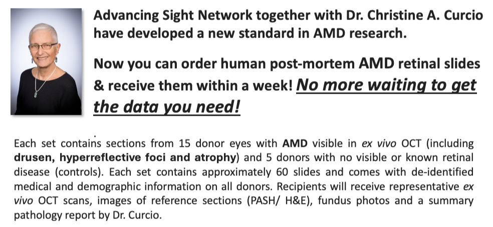
Christine A. Curcio PhD, a neuroscientist by training, has made seminal contributions to the anatomic and molecular pathobiology of age-related macular degeneration (AMD). She was awarded the Proctor Medal (2025), one of the most distinguished honors in ophthalmology research. Using tools of digital histology, her lab has made important discoveries about the composition and role of drusen deposits in human AMD while also identifying hallmarks such as early loss of rod photoreceptors, gliosis, and RPE transdifferentiation. Her microscopy studies support multiple clinical diagnostic techniques, including optical coherence tomography, autofluorescence, adaptive optics, and rod-mediated dark adaptation.
